What is a Chiari Malformation?
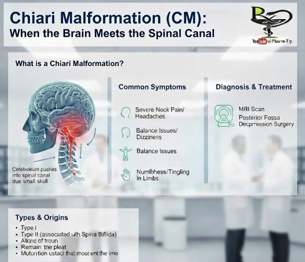
A Chiari Malformation (CM) is a structural abnormality where the lower part of the brain—specifically the cerebellum—is pushed downward through the opening at the base of the skull (the foramen magnum) and into the spinal canal.
The cerebellum is the region of the brain responsible for controlling balance, coordination, and complex motor functions. This displacement typically occurs because the back of the skull is abnormally small or misshapen, creating pressure that forces brain tissue out of its natural position.
Common Symptoms
While some people with CM never experience symptoms (found only during scans for other issues), others may develop symptoms as the pressure on the brainstem and spinal cord increases:
- Pain: Frequent, severe neck pain and “tussive” headaches (triggered by coughing or straining).
- Sensory Issues: Numbness, tingling, or “pins and needles” in the hands and feet.
- Coordination: Balance problems, dizziness, and poor hand-eye coordination.
- Vital Functions: Difficulty swallowing (dysphagia), hoarseness, or vision changes (blurred or double vision).
Types and Origins
There are several classifications of CM, categorized by which parts of the brain are displaced and whether other birth defects are present:
- Type I: The most common form; often doesn’t show symptoms until adolescence or adulthood. It involves the cerebellar tonsils extending into the spinal canal.
- Type II: Also known as “Classic” Chiari Malformation. It is usually associated with Spina Bifida and is typically diagnosed in infants or children.
- Type III and IV: These are much rarer and involve more severe displacement or incomplete development of the cerebellum.
Diagnosis and Treatment
If a doctor suspects CM based on physical symptoms and a neurological exam, they will use imaging to confirm the diagnosis:
- MRI (Magnetic Resonance Imaging): This is the definitive tool for visualizing brain tissue and the extent of the malformation.
- Cine MRI: Used to look at the flow of cerebrospinal fluid (CSF) around the brain and spine.
Management Options
- Observation: If no symptoms are present, doctors may simply monitor the condition with regular scans.
- Medication: Used to manage specific symptoms like pain or headaches.
- Surgery: If symptoms are severe or worsening, a posterior fossa decompression may be performed. The surgeon removes a small piece of bone at the back of the skull to create more room for the brain and restore the normal flow of spinal fluid.