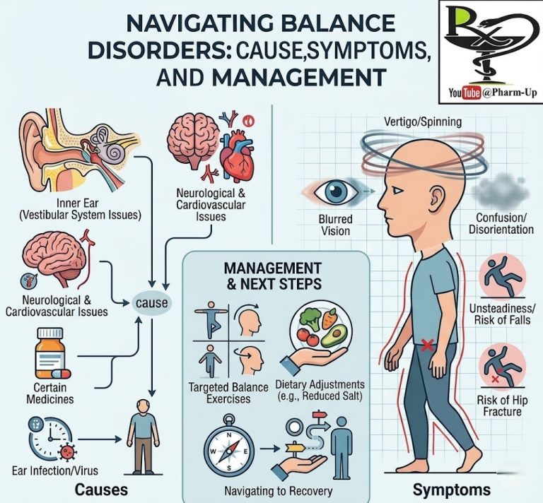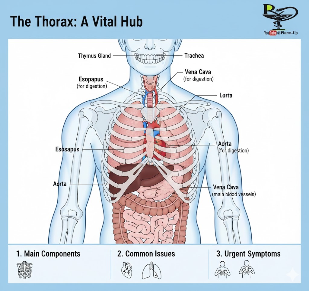
The chest, or thorax, is the region of the body located between the base of the neck and the diaphragm. It serves as a protective cage for the body’s most vital life-support systems. The chest is designed to be both rigid enough to protect the heart and lungs, yet flexible enough to expand and contract during respiration.
1. Key Structures of the Thorax
The chest is a densely packed cavity containing organs and conduits for air, food, and blood.
- The Thoracic Cage: Comprised of 12 pairs of ribs, the sternum (breastbone), and the thoracic spine. It provides structural support and protection.
- The Respiratory System: Includes the trachea (windpipe), which branches into the bronchi, leading into the lungs.
- The Circulatory System: Centered around the heart and the great vessels, such as the aorta (the body’s largest artery) and the vena cava.
- The Digestive Conduit: The esophagus runs behind the trachea to deliver food to the stomach.
- The Pleura: A dual-layered serous membrane that creates a vacuum-like seal around the lungs, allowing them to expand without friction.
2. Common Injuries and Disorders
Because so many systems intersect in the thorax, disorders can range from musculoskeletal issues to life-threatening internal failures.
- Traumatic Injuries:
- Rib Fractures: Can range from simple cracks to “flail chest,” where a segment of the rib cage breaks away.
- Pneumothorax (Collapsed Lung): Occurs when air leaks into the pleural space, putting pressure on the lung and preventing it from inflating.
- Vascular and Cardiac Disorders:
- Thoracic Aortic Aneurysm: A dangerous bulging in the part of the aorta that passes through the chest.
- Heart Disease: Including coronary artery disease or pericarditis (inflammation of the heart sac).
- Mediastinal Diseases: These affect the mediastinum, the central compartment between the lungs that houses the heart, thymus gland, and many lymph nodes.
3. How Chest Problems are Diagnosed
Diagnosis typically begins with a physical exam and a review of symptoms like chest pain, shortness of breath, or difficulty swallowing. Because the chest is opaque, imaging and functional tests are essential.
- Imaging Tests:
- Chest X-ray: The first line of defense to check for broken bones, lung infections, or an enlarged heart.
- CT Scan (Computed Tomography): Provides highly detailed cross-sectional views of blood vessels and soft tissues.
- Ultrasound (Echocardiogram): Specifically used to visualize the heart’s valves and pumping strength.
- Functional Tests:
- Electrocardiogram (ECG/EKG): Measures the electrical activity of the heart.
- Pulmonary Function Tests (PFTs): Measure how well the lungs move air in and out.
- Endoscopy: Using a camera to look inside the esophagus (Upper GI Endoscopy) or the airways (Bronchoscopy).
4. Warning Signs: When to Seek Urgent Care
Chest pain should always be taken seriously. Seek emergency medical help if you experience:
- Sudden, crushing pain or pressure in the center of the chest.
- Pain that radiates to the jaw, neck, or left arm.
- Difficulty breathing or rapid, shallow breathing.
- Coughing up blood.