Because heart disease is the leading cause of death in the United States, early detection is vital. Since the heart is a complex organ with both mechanical (pumping) and electrical (rhythm) systems, doctors use a variety of specialized tests to identify problems before they become life-threatening.
Electrical and Sound Testing (Non-Invasive)
These tests are typically the first step in a cardiac evaluation because they are painless and do not require entering the body.
- Electrocardiogram (EKG/ECG): Records the electrical activity of the heart. It identifies arrhythmias (irregular rhythms) or signs of a past or current heart attack.
- Echocardiography (Echo): Uses ultrasound waves to create moving images of the heart. It allows doctors to see the valves opening and closing and measure the strength of the heart’s pumping action (ejection fraction).
- Stress Testing: Measures how the heart handles physical exertion. By walking on a treadmill while monitored, doctors can see if the heart gets enough blood flow when it is working its hardest.
Advanced Imaging (Detailed Visualization)
If initial tests suggest an issue, advanced imaging provides a 3D view of the heart’s anatomy.
- Cardiac CT Scan: Uses X-rays to create a 3D model of the heart. It is excellent for finding calcium buildup or blockages in the coronary arteries.
- Cardiac MRI: Uses magnets and radio waves (no radiation) to create highly detailed images. It is the “gold standard” for looking at heart muscle damage from a heart attack or identifying cardiac tumors.
- Chest X-Ray: A basic image that shows the overall size of the heart and whether there is fluid in the lungs, which is a key sign of heart failure.
Interventional Procedures (Inside the Heart)
Sometimes, doctors need to go inside the blood vessels to get the most accurate information or to provide immediate treatment.
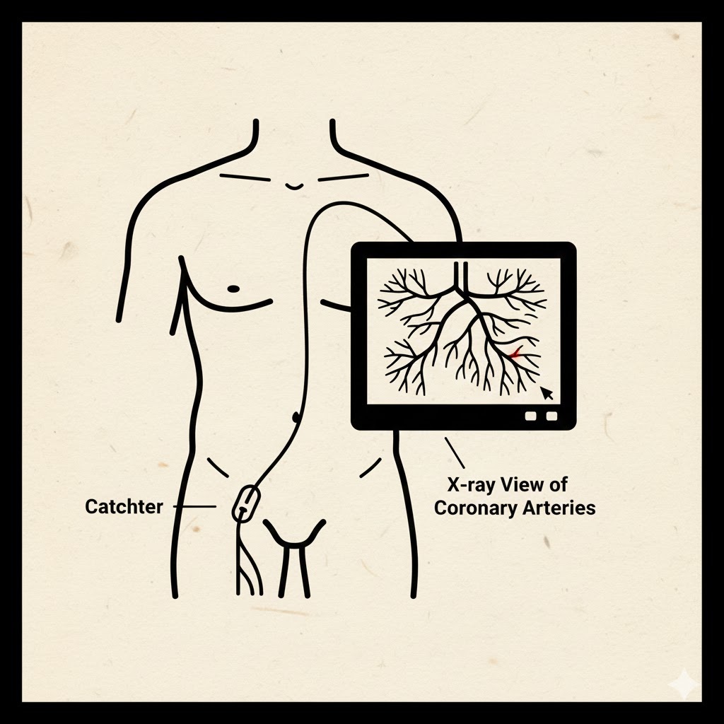
Cardiac Catheterization & Angiography
A long, thin tube (catheter) is threaded through a blood vessel in the arm or groin up to the heart.
- Angiography: A special dye is injected through the catheter. Under X-ray, this dye highlights exactly where and how severely an artery is blocked.
- Treatment: While the catheter is already in place, doctors can often perform an angioplasty (opening a blocked artery with a balloon) or place a stent to keep the artery open.
🔬 Comparison of Common Heart Tests
| Test | Best Used For | Method |
| EKG | Arrhythmias / Heart Attack | Electrical sensors on skin |
| Echo | Valve issues / Pumping strength | Ultrasound (Sound waves) |
| CT Scan | Plaque & Calcium buildup | X-rays / 3D Modeling |
| Stress Test | Clogged arteries during exercise | Treadmill + Monitoring |
| Angiogram | Precise location of blockages | Catheter + Contrast Dye |