New Breakthrough in High-Resolution 3D Body Mapping Revolutionizes Anatomical Education
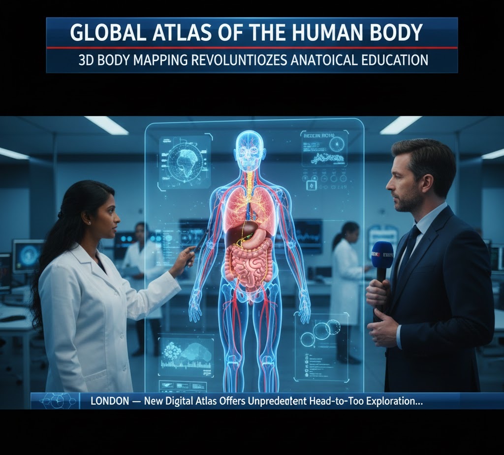
A consortium of medical researchers and software engineers has unveiled the “Global Atlas of the Human Body,” a groundbreaking digital anatomy project that allows users to explore the human form from the cellular level to complete organ systems with unprecedented 3D clarity.
The project, which took five years to complete, aims to replace traditional static diagrams with interactive, “living” models. By integrating thousands of high-resolution MRI and CT scans, the atlas provides a “head-to-toe” perspective that allows medical students and curious laypeople alike to peel back layers of tissue, observe blood flow in real-time, and study the intricate connections between the nervous and muscular systems.
“Anatomy is no longer just about memorizing parts in a textbook,” says Dr. Elena Vance, lead researcher on the project. “It’s about understanding the dynamic relationships between systems. Seeing how the respiratory system interacts with the circulatory system in a 360-degree digital space changes how we teach surgery and diagnose disease.”
The platform is expected to be rolled out to medical schools globally by late 2026, offering a new standard for anatomical accuracy and accessibility.