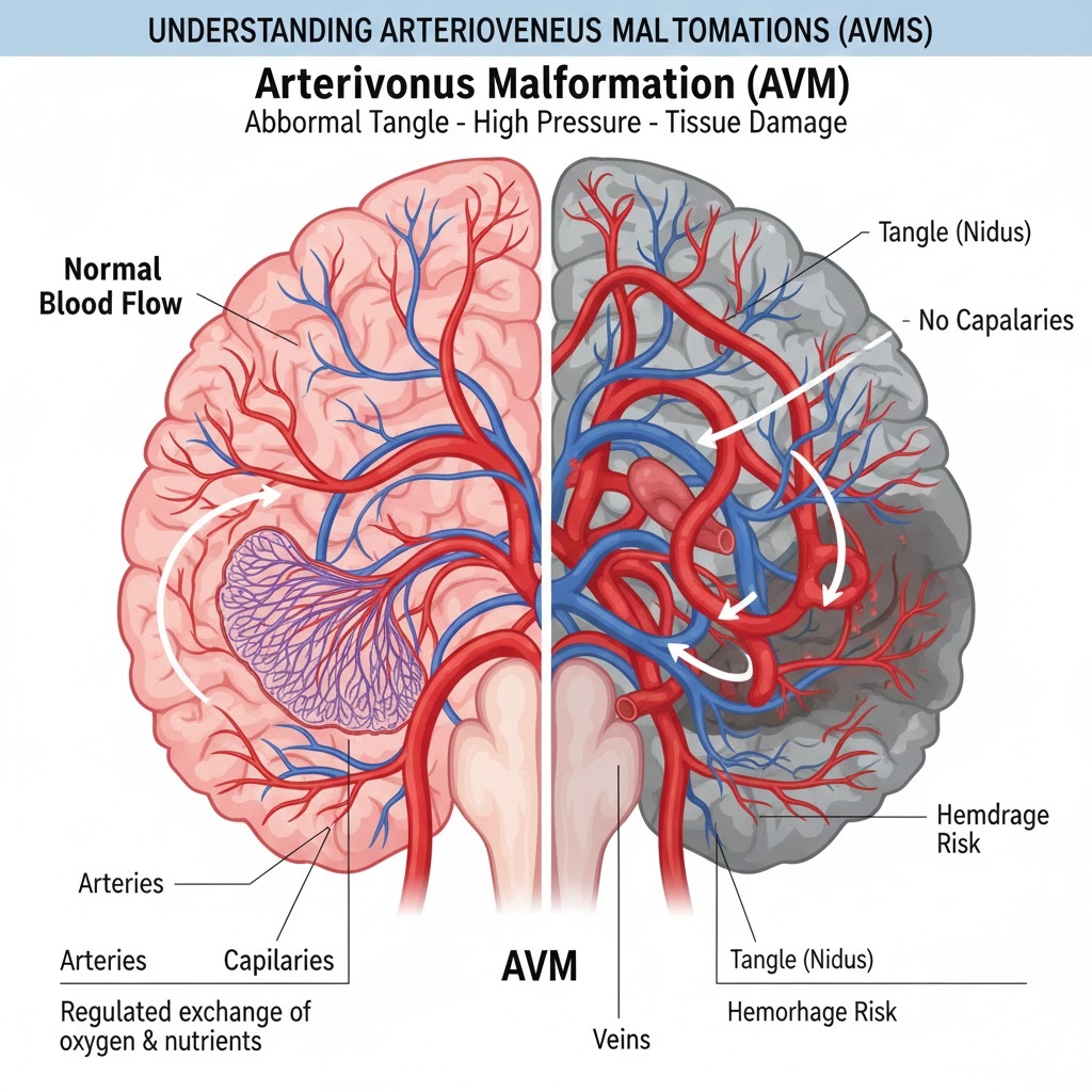
An Arteriovenous Malformation (AVM) is a structural defect in the vascular system characterized by an abnormal “tangle” of blood vessels. To understand why this is dangerous, it helps to look at how blood normally flows through your body.
1. Normal Blood Flow vs. AVM
In a healthy vascular system, blood moves through a specific sequence:
- Arteries: Carry high-pressure, oxygen-rich blood away from the heart.
- Capillaries: Tiny vessels that slow down blood flow, allowing oxygen to move into tissues and waste products to move out.
- Veins: Carry low-pressure, oxygen-depleted blood back to the heart.
In an AVM, the capillaries are missing. The arteries connect directly to the veins. This creates two major problems:
- Tissue Starvation: Because there are no capillaries to distribute oxygen, the surrounding tissue may become damaged or die.
- High Pressure: Veins are not built to handle the high-pressure blood that usually stays in the arteries. This pressure can cause the vessels to stretch, weaken, and potentially hemorrhage (burst).
2. Symptoms and Locations
While AVMs can occur anywhere in the body, they are most clinically significant when located in the brain or spinal cord. Symptoms vary based on location:
- Neurological Symptoms: Seizures, severe headaches, or dizziness.
- Physical Deficits: Muscle weakness, numbness, or problems with vision and speech.
- Cognitive Issues: Confusion or loss of consciousness.
If a brain AVM ruptures, it leads to a hemorrhagic stroke, which is a medical emergency.
3. Diagnosis and Treatment
Diagnosis
Doctors often find AVMs during imaging for unrelated issues, or after a patient reports symptoms. Common tests include:
- MRI/CT Scans: To see the structure of the brain or spine.
- Cerebral Angiography: The “gold standard” for diagnosis, where dye is injected to map the blood flow exactly.
- Bruit Check: A doctor may use a stethoscope to listen for a “whooshing” sound (bruit) caused by the rapid blood flow.
Treatment Options
Treatment is highly individualized based on the risk of rupture:
- Medical Management: Using drugs to control seizures or headaches.
- Endovascular Embolization: A catheter is used to “plug” the abnormal vessels with a glue-like substance to reduce blood flow.
- Stereotactic Radiosurgery: Using focused radiation to slowly scar and close the AVM over time.
- Surgical Resection: Physically removing the tangle of vessels if it is in a reachable area of the brain.