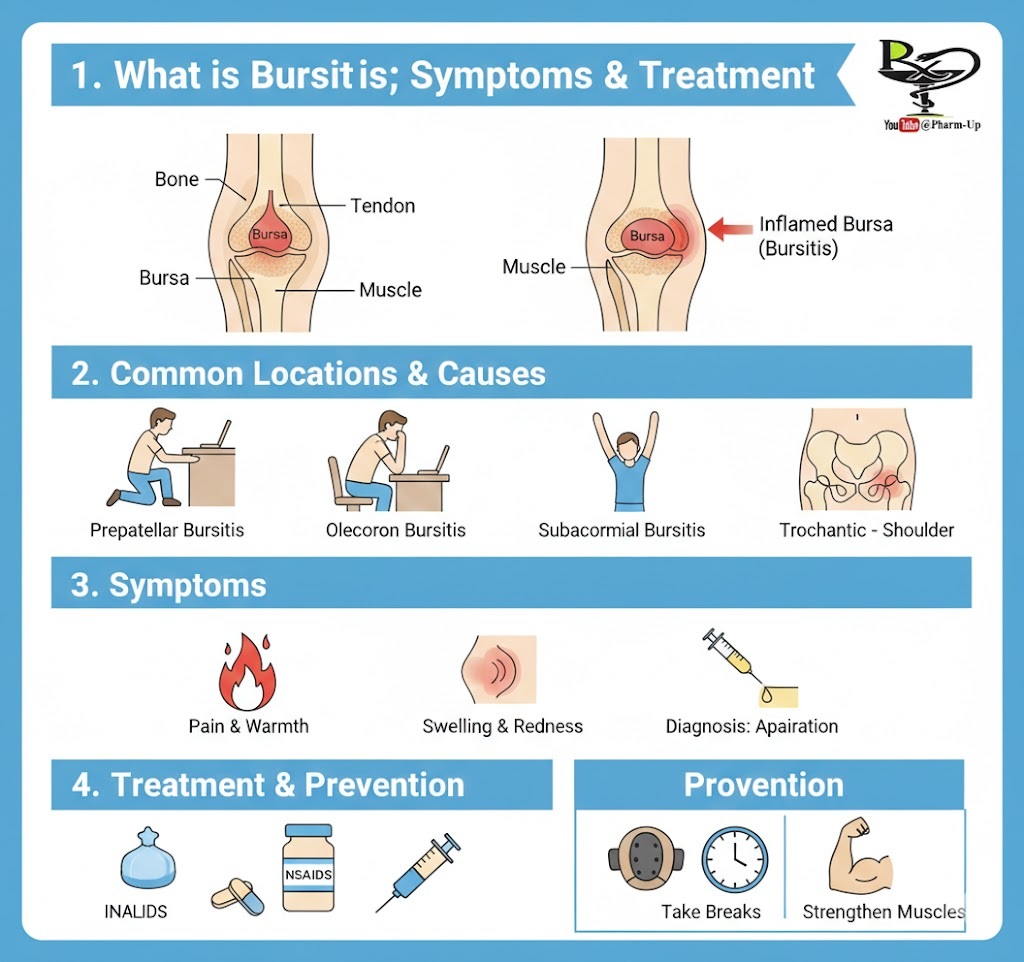
A bursa (plural: bursae) is a tiny, slippery sac filled with synovial fluid. These sacs serve a vital purpose: they act as biological “cushions” or “lubricants” between hard bone and softer tissues like tendons and muscles, preventing painful friction. When these sacs become inflamed, the condition is known as bursitis.
1. How Bursitis Develops
Bursitis is primarily an “overuse” injury, though it can stem from several sources:
- Repetitive Stress: Making the same motion repeatedly (e.g., pitching a baseball, lifting overhead, or even typing) can irritate the bursa.
- Prolonged Pressure: Constant leaning on the elbows (“Student’s Elbow”) or kneeling on hard floors (“Housemaid’s Knee”) puts direct pressure on the sac.
- Acute Injury: A sudden blow or fall directly onto a joint can cause the bursa to fill with excess fluid or blood.
- Age and Occupation: As we age, tendons become less flexible and the risk of bursitis increases. Occupations like carpentry, gardening, or painting carry higher risks.
2. Common Locations of Bursitis
While there are over 150 bursae in the human body, bursitis most commonly occurs in joints with a high range of motion:
- Elbow (Olecranon): Often caused by leaning on hard desks.
- Knee (Prepatellar): Common in people who kneel frequently.
- Shoulder (Subacromial): Linked to reaching overhead.
- Hip (Trochanteric): Often causes pain when lying on one’s side.
3. Symptoms and Diagnosis
The primary signs of bursitis include:
- Pain: Usually a dull ache or stiffness that worsens with movement or pressure.
- Visible Swelling: The joint may look “puffy” or have a distinct lump.
- Redness and Warmth: If the area is hot to the touch, it may indicate septic bursitis (an infection), which is a medical emergency.
Diagnostic Steps:
Doctors typically use a physical exam to check for tenderness. If the diagnosis is unclear, they may order:
- Imaging: X-rays (to rule out bone issues) or MRIs.
- Aspiration: Using a needle to draw fluid from the bursa to test for bacteria (infection) or gout crystals.
4. Treatment and Recovery Pathways
Most cases of bursitis can be managed at home, but chronic cases require more intensive care.
| Treatment Level | Methods Used |
| Stage 1: Conservative | R.I.C.E. (Rest, Ice, Compression, Elevation) and NSAIDs like ibuprofen. |
| Stage 2: Clinical | Corticosteroid Injections to quickly reduce inflammation or physical therapy to strengthen supporting muscles. |
| Stage 3: Advanced | Drainage of the fluid or, in rare cases (6-12 months of no improvement), Bursectomy (surgical removal of the bursa). |
5. Prevention Tips
- Use Padding: Wear knee pads when gardening or elbow pads when leaning.
- Take Breaks: If you perform repetitive tasks, set a timer to rest your joints every 20 minutes.
- Strengthen Muscles: Stronger muscles help share the load, taking pressure off the bursa.